New customers save 5% with the code GET05




Human Anatomy Models in India | Esaw India
by Akash Dhawan
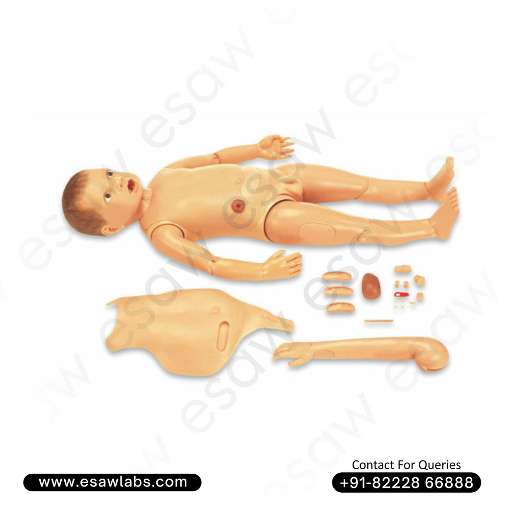
The human body is an architectural masterpiece of biological engineering. From the microscopic pathways of neural networks to the macro-structures of musculoskeletal systems, understanding how these components interact is the cornerstone of all medical, nursing, and life science education. While digital simulation software and virtual reality anatomy tools have made significant technological leaps, tactile, three-dimensional physical representations remain irreplaceable. For medical universities, nursing institutes, biology labs, and healthcare training centers, navigating the procurement landscape for these educational tools requires finding top-tier Human Anatomy Models in India.
As medical curricula evolve to emphasize highly practical, hands-on diagnostic training, the demand for dimensionally accurate, highly durable, and realistic anatomical replicas has scaled rapidly. Esaw India, a pioneering force in the scientific instrumentation and educational supply sector since 1962, manufactures and supplies medical-grade structural representations that map human physiology with absolute fidelity. This comprehensive informational guide deep-dives into the scientific necessity of three-dimensional anatomical tracking, the engineering standards that define high-quality production, and what institutional buyers must evaluate when sourcing these critical assets.
The Educational Necessity of 3D Human Anatomy Models
Textbooks and two-dimensional medical diagrams offer conceptual frameworks, but they fall short in conveying spatial awareness. A medical student cannot truly grasp the depth, spatial orientation, or physical relationships of organs inside the thoracic cavity from a flat screen or page. High-fidelity Human Anatomy Models in India solve this structural visualization gap.
Spatial Awareness and Deep Learning
Medical education relies heavily on understanding physical topology—knowing exactly how an artery loops around a bone or how a muscle insertion relates to a nerve bundle. Three-dimensional models allow students to physically interact with structures, rotate them, disassemble interlocking organ modules, and visually trace physiological pathways. This tactile exploration bridges the gap between theoretical knowledge and practical surgical or clinical execution.
Standardizing Medical Training across Institutional Tiers
Not every medical college or nursing school has immediate, unrestricted access to cadaveric dissection labs due to high maintenance costs, legal constraints, and ethical procurement challenges. High-quality Human Anatomy Models in India serve as a vital, standardized alternative. They ensure that students across all tiers of training can repeatedly examine human structural systems without degradation, tissue decay, or time constraints.
Architectural Taxonomy: Key Categories of Anatomical Replicas
To build a fully functional, comprehensive medical training or biological laboratory, institutions must deploy a diverse inventory of structural models. Professional suppliers organize these models into distinct taxonomic categories based on medical specialization.
1. Osteological Replicas (Full Skeletons and Bone Structures)
The skeletal framework is the foundation of anatomical study. High-grade osteological models must represent realistic bone texture, weight distribution, and precise articulation points:
-
Life-Sized Articulated Skeletons: Mounted on roller stands, these replicas display all 206 human bones, complete with realistic skull suturing, nerve branches, and vertebral artery pathways.
-
Disarticulated Skeletons: A complete set of unjoined bones, allowing students to inspect individual skull plates, ribs, vertebrae, and phalanges in isolation.
-
Functional Joint Models: Highly detailed representations of the shoulder, hip, knee, and elbow joints, incorporating flexible synthetic ligaments to demonstrate natural biomechanical movement, extension, flexion, and rotation.
2. Dissectible Torso Models
The ultimate centerpiece of any biology or nursing lab is the multi-part human torso model. Designed for deep internal organ analysis, these models typically feature removable components:
-
Interchangeable Genital Inserts: Dual-sex configurations that allow institutions to swap male and female reproductive systems within a single torso framework, optimizing budget utilization.
-
Sectioned Internal Organs: Removable lung halves, disassemblable two or four-part hearts, stomach sections, and exposed intestinal tracts that clearly map out the vascular and digestive topography.
3. Specialized Systemic and Organ Models
For advanced clinical tracks like neurology, cardiology, ophthalmology, and dentistry, highly magnified organ replicas are essential:
-
Neuro-Anatomy Replicas: Multiple-part brain models sectioned along median planes to reveal internal structures like the ventricles, cerebellum, brainstem, and pituitary gland.
-
Cardiovascular Modules: Giant-sized heart models displaying internal valve structures, coronary arteries, and systemic circulatory loops.
-
Ophthalmology and Otology Tools: Magnified eye and ear models that disassemble to show retinal layers, auditory ossicles, and cochlear structures.
Material Engineering: How Medical Replicas Are Crafted for Longevity
When selecting Human Anatomy Models in India, looking beyond surface aesthetics to understand the chemical composition and manufacturing processes of the materials is critical.
Premium Polyvinyl Chloride (PVC) Construction
The gold standard for industrial-grade anatomical models is high-impact, medical-grade Polyvinyl Chloride (PVC). This synthetic polymer is deliberately selected for several vital chemical properties:
-
Thermal and Environmental Stability: PVC does not warp, melt, or lose its structural form under varying laboratory temperatures or high humidity levels.
-
Chemical Resistance: Models molded from high-grade PVC can be routinely sanitized, wiped down, or cleaned with alcohol-based disinfectants to prevent cross-contamination without degrading the material or fading the paint.
-
Drop and Impact Resistance: Educational settings are high-wear environments. PVC ensures that models can survive accidental drops or rigorous handling by hundreds of students every semester.
Advanced Painting, Texturing, and Color-Coding
A model is only as useful as its visibility. Premium models utilize non-toxic, eco-friendly pigments that are deeply bonded to the plastic material to prevent chipping, flaking, or fading over decades of exposure to ambient light.
-
Systemic Color Standardization: Arteries are universally painted vivid red, veins deep blue, nerves bright yellow, and lymphatic vessels green. This uniform color-coding drastically simplifies structural tracing for beginner students.
-
Hand-Painted Precision: Top manufacturers employ skilled artisans to hand-paint the minute fine lines, striations, and boundaries of muscle fibers and small vascular branches, providing a layer of definition that automated industrial painting cannot match.
Procurement Checkpoints: Selecting the Best Sourcing Partner
Institutions allocating capital budgets for laboratory infrastructure must treat procurement as a long-term investment. Sourcing directly from an established domestic manufacturer-supplier like Esaw India yields distinct operational advantages:
Hand-to-Eye Calibration and Scale Accuracy
Anatomical models must strictly adhere to human scales and proportional dimensions. Inaccurate modeling—such as disproportionate organ sizes or misplaced nerve pathways—can inadvertently teach students wrong physiological associations. Partnering with a specialized supplier ensures that all molds are cast from real human specimens or mathematically precise digital medical scans.
Seamless Part Replacement Protocols
Anatomical torso models contain dozens of small, interlocking parts, pins, and magnetic keys. Over years of student use, minor components like single teeth, specific heart valves, or kidney cross-sections can occasionally be misplaced. Sourcing from a factory-direct provider allows your institution to order exact component replacements without needing to discard the entire base model.
Conclusion: Building a Vision for Advanced Medical Training
As global healthcare standards rise, the training infrastructure inside Indian educational labs must keep pace. High-fidelity physical replicas act as the perfect gateway for young minds to discover the inner workings of human existence. Esaw India masterfully blends decades of engineering heritage with meticulous artistic craftsmanship to stand out as a trusted supplier of Human Anatomy Models in India. By delivering absolute structural accuracy, structural durability, and vivid clarity, we ensure that your laboratory remains a sanctuary for accurate, lifelong learning.
Equip your lecture halls, training labs, and biological institutes with instruments designed for maximum structural longevity and uncompromised precision. Partner with Esaw India and bring absolute clarity to the science of human anatomy.
Tag:Best Human Anatomy ModelsHuman Anatomy ModelHuman Anatomy ModelsHuman Anatomy Models DealerHuman Anatomy Models ExporterHuman Anatomy Models in IndiaHuman Anatomy Models ManufacturerHuman Anatomy Models supplierHuman Anatomy Models WholsalerThe Human Anatomy Models in IndiaTop 10 Human Anatomy ModelsTop 10 Human Anatomy Models in IndiaTop 5 Human Anatomy Models




Leave a comment